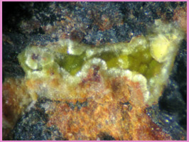
Locality: Ham-Weeks Quarry, Wakefield, NH
Specimen Size: 3 mm field of view
Field Collected: Bob Janules. A Bob Janules collection specimen
Catalog No.:
Notes: Identified by EDS analysis. The Al peak in the plot is thought to be due to electron scattering from inside the SEM chamber. The analyst stated: "Uranium does a very good job of scattering the beam."

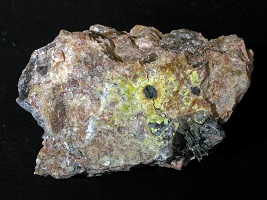
Locality: Ham-Weeks Quarry, Wakefield, NH
Specimen Size: 5 cm specimen. Uranophane aureole surrounding black uraninite bleb.
Field Collected: Tom Mortimer - Aug, 2011
Catalog No.: 1796
Notes:

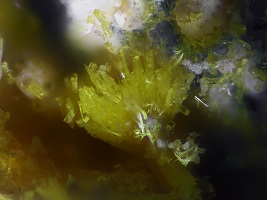
Locality: Ruggles Mine, Grafton, NH
Specimen Size: 0.4 mm uranophane spray
Field Collected: Harvard research
Catalog No.: u2377
Notes: The few scattered orthorhombic prisms are likely phosphuranylite.
Uranophane and uranophane-beta are chemically equivalent, monoclinic, dimorphs. Presently (2020) these are grouped together as uranophane.

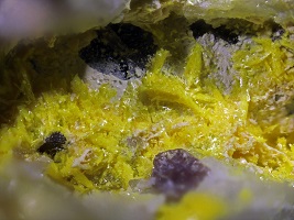
Locality: Ruggles Mine, Grafton, NH
Specimen Size: 4 mm field of view
Field Collected: Harvard research
Catalog No.: u2377
Notes:

Locality: Ruggles Mine, Grafton, NH
Specimen Size: 4 cm specimen
Field Collected: Tom Mortimer - 9/17/21
Catalog No.: 2182
Notes: Yellow crust of uranophane on feldspar. Green arrow points to EDS sample point.
EDS analyses (BC562) indicated a chemistry of Ca1.0U4.6Si2.8O34.6 , normalized for one Ca. The uranium content is very high for uranophane, but still the best species fit. Small amounts of P, Al, Mn, Fe, and Pb suggest that other uranium secondary species may be mixed in.
Uranophane chemistry is Ca(UO2)2[SiO3(OH)]2 · 5H2O .